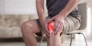
Vein disorders like varicose veins, deep vein thrombosis (DVT), and chronic venous insufficiency can have a substantial impact on an individual’s lifestyle, producing discomfort, pain, and other severe health problems. Contemporary medical imaging provides a non-surgical, highly effective means of diagnosing such conditions. Diagnosing vein problems using ultrasound is a key method physicians and vein specialists in Stroudsburg or vein specialists in Bethlehem use to evaluate vein health and determine appropriate vein treatment strategies. Whether you have noticeable swelling or recurring pain, this imaging technique can give you invaluable information about what’s occurring inside your veins.

What Is a Venous Ultrasound?
A venous ultrasound, also known as a venous duplex scan, employs high-frequency sound waves which take detailed pictures of the veins and quantify blood flow. Unlike X-rays or CT scans, this procedure does not require radiation and is, therefore, a safe and effective choice for vein diagnosis. “Duplex” describes using two forms of ultrasound: one to create detailed pictures of veins and the other (Doppler ultrasound) to measure blood movement within those veins. Venous ultrasound’s capacity to image both the structure of the veins and the flow of blood within them makes it a particularly useful diagnostic tool for detecting problems like blockages, clots, and valve dysfunction. Used either to scan surface veins in the legs or subcutaneous structures, venous ultrasound accurately reflects venous health.
How It Helps Identify Vein Conditions
Vein ultrasound diagnosis offers priceless real-time imaging that assists in detecting various vein conditions. The process provides more than visual imaging; it also helps determine the veins’ functionality by analyzing blood flow through veins. This is how it functions:
Visualising Vein Structure: Ultrasound, which uses high-frequency sound waves, produces detailed images of the veins, delineating blockages, dilations, or abnormalities. Ultrasound for varicose veins is particularly important when evaluating these conditions, as it delineates affected veins and measures their size and structure. Specialists at clinics like vein specialist Stroudsburg and vein specialist Bethlehem often use this method to recommend the most effective vein treatment.
Testing Blood Flow: The Doppler ultrasound lets doctors test how blood flows through the veins. It detects places where blood is not flowing correctly, a characteristic of chronic venous insufficiency or deep vein thrombosis (DVT). The Doppler effect signifies areas of abnormally flowing blood, including venous reflux, where blood flows reverse because of damaged valves.
Detection of Blockages or Clots: Since DVT is a significant risk factor for more serious complications, ultrasound enables physicians to identify blood clots in the veins. It allows them to determine if they are blocking the flow of blood and whether they may develop into life-threatening complications such as pulmonary embolism.
Valve Dysfunction Identification: Ultrasound also evaluates the valve’s function. In cases where valves don’t function well, blood accumulates and forms symptoms such as swelling and heaviness. The procedure identifies such occurrences in the superficial veins and recommends proper intervention.
By presenting this type of detailed, real-time information, ultrasound plays a critical role in diagnosing the apparent symptoms and root causes of vein problems. In doing so, doctors can make better decisions regarding the right treatment options, ranging from lifestyle modifications to more sophisticated medical therapies. Many patients seek vein specialists in East Stroudsburg or vein specialists in Bethlehem to ensure they receive expert guidance and tailored vein treatment solutions.
What to Expect During the Scan
A vein ultrasound diagnosis is brief, simple, and painless. Here is a step-by-step guide to what you can expect during the procedure:
Preparation: The test usually lasts 30 to 60 minutes. You will be asked to lie on a table, and depending on the area under evaluation, you might be required to raise your legs a bit so that better imaging can be achieved.
Applying the Gel: The technician applies a warm, water-like gel to the inspected area to allow the ultrasound waves to pass through your skin efficiently. The gel is not painful and will be wiped off after use.
Using the Ultrasound Probe: A small handheld device, referred to as a transducer, will be placed on the skin. The transducer sends sound waves that reflect off the veins and back to the device, forming images that appear on a monitor.
Doppler Ultrasound: In addition to taking pictures of vein structure, Doppler ultrasound evaluates blood flow. The technician will ask you to move about, change positions, or make gentle movements, such as flexing your ankles or standing up, to test blood flow and identify any abnormal flow or reflux.
Completion: Once the test is over, no recovery time is needed. You may resume your daily routine, which makes the test convenient and easy.
Conditions Typically Diagnosed
As ultrasound assists in determining vein conditions, there are certain health conditions which are especially identified or diagnosed with it. Here are some of the most prevalent conditions that are picked up by the process:
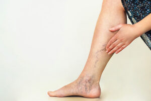
- Varicose Veins
Varicose veins are dilated, engorged veins which usually manifest in the leg. Ultrasound assists physicians in visualizing these veins and determining the degree of damage. By identifying vein dilation and quantifying blood reflux, ultrasound plays a critical role in identifying whether or not conservative therapies such as compression therapy will be adequate or if more aggressive approaches, including sclerotherapy or laser ablation, are indicated. For comprehensive care, patients often consult vein specialists in Stroudsburg for advanced vein treatment options. - Deep Vein Thrombosis (DVT)
DVT is a condition in which a blood clot occurs in the deep veins of the legs. When the clot becomes dislodged and flows to the lungs, it is fatal, leading to pulmonary embolism. Ultrasound application on veins is the most accurate way to detect DVT. It can display the size and location of the clot so that doctors can give the right treatment, which could be anticoagulants or surgery. - Chronic Venous Insufficiency (CVI)
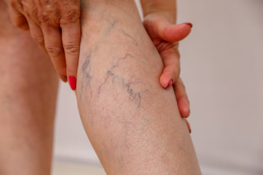
Chronic venous insuffciency occurs when veins are unable to effectively return blood to the heart, usually due to valve dysfunction. This creates symptoms like leg swelling, pain, and changes in the skin. Ultrasound plays a vital role in CVI diagnosis by detecting areas of venous reflux and evaluating vein valve health. The imaging also aids in tracking the disease’s progression and informing treatment choices. - Superficial Thrombophlebitis
This condition is an inflammation of a vein close to the skin’s surface, typically associated with a blood clot. Superficial thrombophlebitis is generally less serious than DVT, but it may cause discomfort and swelling. Ultrasound allows for accurate imaging to distinguish superficial clots from deeper, more dangerous ones. - Venous Malformations
Venous malformations are irregular collections of veins that can cause swelling, pain, and even ulcers. They are usually present at birth (congenital) but do not necessarily occur until adulthood. Ultrasound reveals these venous abnormalities, which leads to the selection of appropriate treatments on time, such as embolisation or surgery. - Post-Thrombotic Syndrome
Post-thrombotic syndrome is a condition that may follow an individual who has experienced a DVT. It may result in chronic pain, swelling, and skin colour change. Ultrasound is employed to assess the affected veins and determine if there are residual clots or valve dysfunction that may be causing recurring symptoms. - Superficial Venous Thrombosis
In contrast to DVT, which occurs deeper in the veins, superficial venous thrombosis comprises clots in more superficial veins. Ultrasound assists physicians in assessing the size of the clot and how it impacts the blood flow through the involved veins. Depending on the level of thrombosis, treatment can be as simple as anti-inflammatory drugs or more sophisticated procedures.
The Role of Imaging in Treatment
Proper imaging is essential to ascertain the appropriate course of vein treatment for vein conditions. Without the appropriate visualization of the veins and blood flow, it may not be easy to choose the most suitable intervention, which could result in ineffective or unnecessary treatment. Vein ultrasound diagnosis gives physicians the transparency they require in order to:
Verify the Diagnosis: Imaging assists in correctly diagnosing the underlying cause of symptoms, whether it’s a clot, valve malfunction, or other reason.
Individualised Treatment Plans: Based on the results, medical practitioners can develop individual treatment plans aimed at specific affected veins via conservative treatments or more complex procedures.
Track Progress: For individuals receiving vein treatment, regular ultrasounds can track the progress of the treatment and identify early signs of possible complications.

Conclusion
In short, vein ultrasound diagnosis is a vital and effective diagnostic tool for diagnosing various venous disorders, from varicose veins to DVT and chronic venous insufficiency. It offers essential real-time information that enables doctors to develop individualized treatment plans and ensures patients receive optimal care. If you have symptoms like leg swelling, pain, or prominent veins, consult a vein specialist Stroudsburg for expert vein treatment. Early diagnosis and treatment are essential in preventing complications and maintaining overall vein health.
FAQ – Vein Ultrasound and Vein Treatment
1. Why should I visit a vein specialist in Stroudsburg or Bethlehem?
Visiting a vein specialist in Stroudsburg or a vein specialist in Easton ensures you receive expert diagnosis and personalized care for your vein concerns. Specialists use advanced imaging techniques like venous ultrasound to detect underlying problems such as varicose veins, DVT, or valve dysfunction early—helping prevent complications and guiding you toward the most effective vein treatment options.
2. Is a venous ultrasound painful?
No, a venous ultrasound is completely painless and non-invasive. It involves applying a warm gel and gently moving a small handheld device (transducer) over your skin to capture images of your veins. You can resume normal activities immediately after the test.
3. How long does a vein ultrasound take?
Typically, a venous ultrasound takes between 30 to 60 minutes, depending on the area being examined. The process is quick, comfortable, and doesn’t require any recovery time.
4. What conditions can a vein ultrasound detect?
A vein ultrasound can identify several vein conditions, including varicose veins, deep vein thrombosis (DVT), chronic venous insufficiency (CVI), superficial thrombophlebitis, and venous malformations. It also helps specialists determine the severity of the condition and plan the right vein treatment.
5. How should I prepare for a venous ultrasound?
There’s no special preparation needed. You may be advised to wear loose-fitting clothing and avoid applying lotions to your legs before the scan. During the test, the technician may ask you to change positions or move slightly to assess blood flow properly.
6. What are the benefits of getting a venous ultrasound?
A venous ultrasound provides real-time imaging that helps identify blockages, valve dysfunction, or abnormal blood flow. It enables your doctor or vein specialist to recommend targeted vein treatments, ranging from lifestyle changes and compression therapy to minimally invasive procedures like sclerotherapy or ablation.
7. How soon will I get my results?
In most cases, results from a venous ultrasound are available immediately after the scan or within a short time. The vein specialist will review your images, discuss findings, and suggest appropriate next steps for your vein treatment plan.
8. When should I consider seeing a vein specialist?
You should visit a vein specialist in Stroudsburg or a vein specialist in Bethlehem if you experience symptoms like leg swelling, heaviness, visible varicose veins, skin discoloration, or persistent pain. Early evaluation can prevent progression and help you maintain healthy circulation.
9. Can venous ultrasound be used to monitor treatment progress?
Yes, ultrasound imaging is an excellent way to track the effectiveness of your vein treatment over time. It helps specialists monitor improvements, detect early signs of complications, and make any necessary adjustments to your care plan.
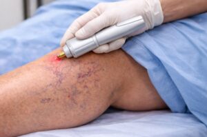
10. Are vein treatments safe and effective?
Yes, modern vein treatments are safe, minimally invasive, and highly effective. Procedures like laser therapy, sclerotherapy, and radiofrequency ablation offer fast recovery times and excellent long-term results, especially when guided by experienced specialists.